

IMAN HASSANI
PH.D. CANDIDATE, DEPARTMENT OF CHEMICAL ENGINEERING, COLLEGE OF SCIENCES AND MATHEMATICS
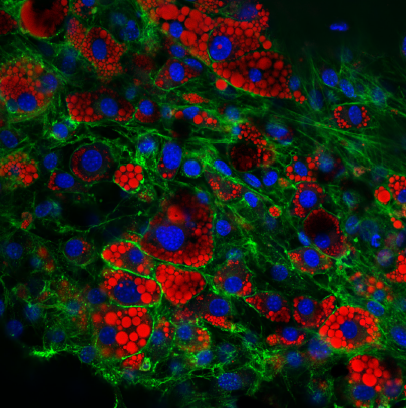
ELUCIDATION OF THE ADIPOCYTIC MICROENVIRONMENT:
To study the impact of fatty tissue on the incidence and progression of colorectal cancer, 3T3-L1 mouse fibroblast cells were differentiated to express an adipocytic, or fat cell, phenotype. To visualize cellular morphology, the adipocytes were stained with Hoechst 33342, WGA, and Oil Red O to illuminate cellular nuclei in blue, plasma membrane in green, and lipid droplets in red, respectively. The cells were then imaged utilizing confocal microscopy. When printed at 28”x 28”, this image elucidates the cellular microenvironment at 2,235 times larger than its actual size.

Biology, Science Photography
Confocal microscopic image of fat cells
28” x 28”
2018
Last Updated: July 24, 2018